
X-RAY DIAGNOSIS AND ANALYSIS
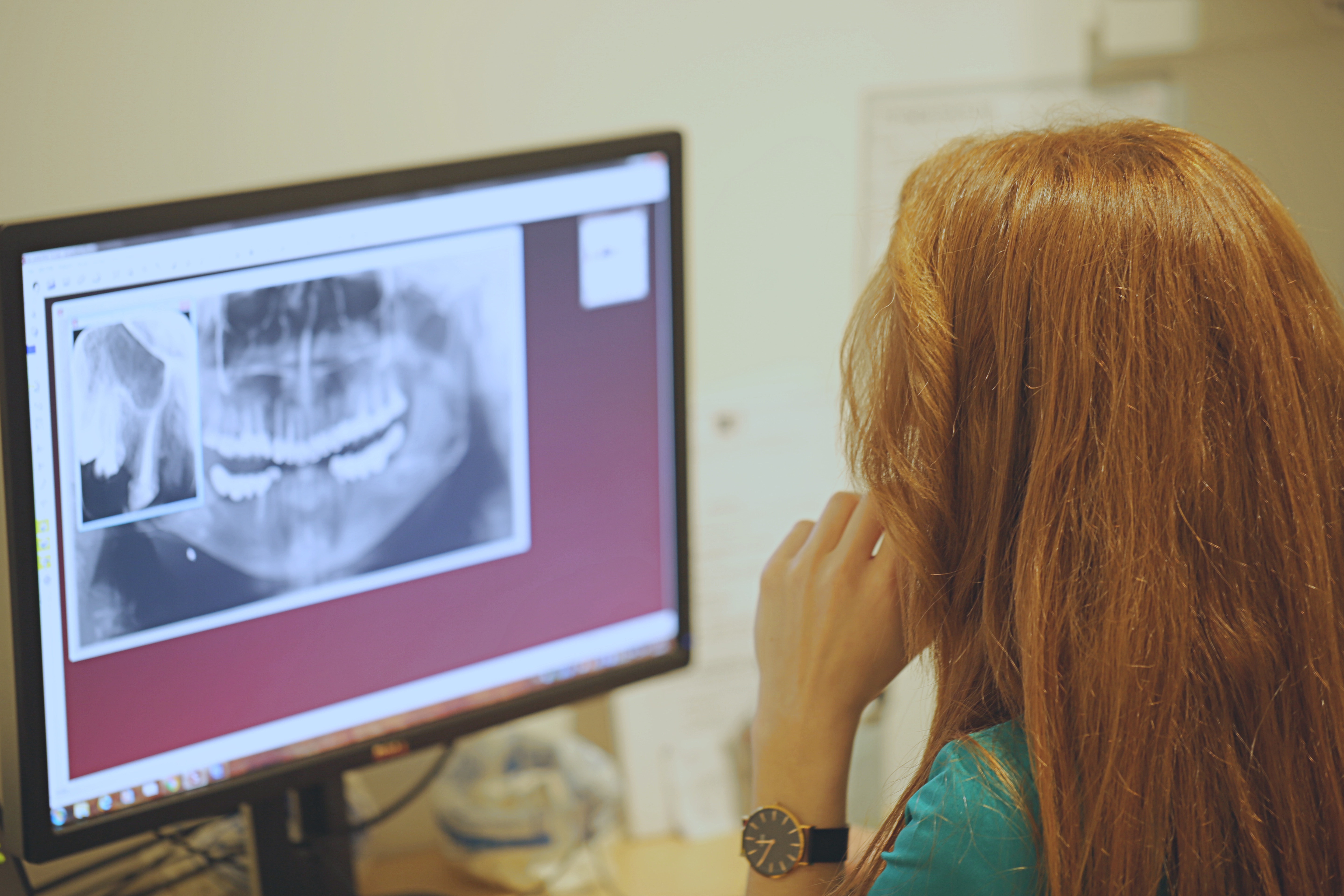
Before each therapy, an analysis and diagnosis of the patient's oral status is performed. We start the diagnosis with a dental examination of the patient's oral cavity using an intraoral camera, and then with an X-ray. In our clinic we use the Soredex CRANEX 3D X-ray device, which is the most modern digital X-ray system on the market and emits extremely small doses of radiation.
Depending on the situation and the required analysis, we record the patient:
- Small intraoral RVG images
- 2D panoramic images (Orthopantomogram)
- 3D CBCT layered images of teeth and jaws
1. What are the advantages of small intraoral RVG images of teeth
The advantage of small intraoral RVG images of teeth is that the images are of exceptional quality and are available to our doctors on a monitor via a digital sensor in just a few moments. Small intraoral RVG images are single images of teeth that are done when the doctor applies therapy to a narrower area of the patient's jaw, and there is a need for a very detailed image of the teeth at high magnification.
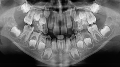
2. What are the advantages of 2D panoramic images (orthopantomogram)?
The advantage of 2D panoramic images is a high-quality X-ray display of both jaws without opening the mouth, with a small amount of radiation, while the procedure itself is not complicated. We permanently archive all recordings of our patients in our system.
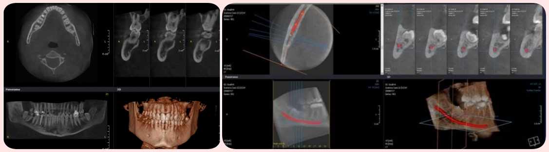
3. What are the benefits of 3D CBCT layered imaging of teeth and jaws?
The advantage of CT computed tomography is in the stratification of the image. With the help of 3D CT images, we get a view of the stratification of the bone, which can be up to 0.2 millimetres, and thus there is no possibility that we miss any information in the analysis.
4. What X-rays are needed in restorative dentistry?
X-rays required in conservative dentistry are a small intraoral RVG image or a 2D panoramic image (orthopantomogram), depending on the area of therapy required. If we are doing individual tooth positions imaging, then we recommend a small intraoral image (RVG), and if it is a larger number of individual teeth at different positions, then we recommend a 2D panoramic image (orthopantomogram) to patients. Both types of images are fast and of exceptional quality, and the patient can take them during the examination, without the need to leave the clinic, and completely free of charge.
5. What X-rays are needed for surgical and implant therapy?
For the purpose of successful planning of surgical and implant therapy, we use 3D CBCT images. Their biggest advantage is the high-quality diagnostic data necessary in the diagnosis of various diseases of the teeth, jaws and surrounding structures. With the help of 3D CT images, we get layered images of teeth and jaws, which are the most important item in the protocol for planning and performing surgical or implant therapy. Nowadays, the most common mistake in dentistry is to plan implant therapy based on a 2D X-ray (orthopantomogram), which cannot show the current condition and volume of the patient's bone. For example, if a patient is fitted with implants based on a 2D X-ray, there is a high possibility that the patient's jawbone does not have sufficient volume or strength to osseointegrate the implant, which results in repetition of all work without wasting patient time and money. Display of the strength and volume of the patient's bone is only possible with 3D CBCT images, and any surgical or implant therapy must be planned with the help of the mentioned images!
Contact Us


Menu
Home